Author : Alicia Harlov
Alicia Harlov is one of Flex Boots’ Equine Educators and Barefoot Trimming Specialists, where she creates video content as well as written articles pertinent to barefoot hoof health and soundness. Outside of Flex Boots, Alicia is a PHCP-certified hoofcare provider and program mentor who lives on the North Shore of Massachusetts, where she runs a hoof rehabilitation facility with a special focus on navicular horses. She pursues continuing education for hoofcare, equine nutrition and biomechanics at every opportunity, from sources around the world.
Radiographs can be an incredibly useful tool when we’re trying to understand what is happening inside a horse’s hoof. They give us information that we simply can’t get by looking at the outside of the foot. But as useful as radiographs are, they are only part of the picture.
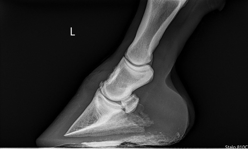
We can look at a radiograph and identify changes in bone, measure relationships within the hoof capsule, and sometimes discover abnormalities we never would have known were there. What we can’t necessarily do is look at that image and say, “There. That’s why this horse hurts.” We also can’t look at a radiograph in isolation and have it tell us exactly how that individual horse’s foot should be trimmed. Understanding both the value and the limitations of radiographs can help us use them much more effectively.
What Can Radiographs Tell Us?
Radiographs are particularly useful for looking at bone, gas pockets, and things like dense foreign objects. Within the hoof, we can evaluate structures such as the coffin bone, navicular bone, and sometimes portions of the pastern bones. Depending on the views taken and the quality of the images, radiographs can help identify fractures, bone remodeling, areas of bone loss or proliferation, changes involving the navicular bone, mineralization, and other abnormalities.
With appropriately positioned and marked hoof radiographs, we can also evaluate relationships within the hoof that aren’t always obvious from the outside. We can look at things like sole depth beneath the coffin bone, the orientation of the coffin bone relative to the ground, the relationship between the dorsal hoof wall and the coffin bone, phalangeal alignment, and changes associated with laminitis. This can give both the veterinarian and hoofcare provider valuable information about what’s happening internally.
But we’re still looking primarily at a snapshot of anatomy. We’re not looking directly at pain.
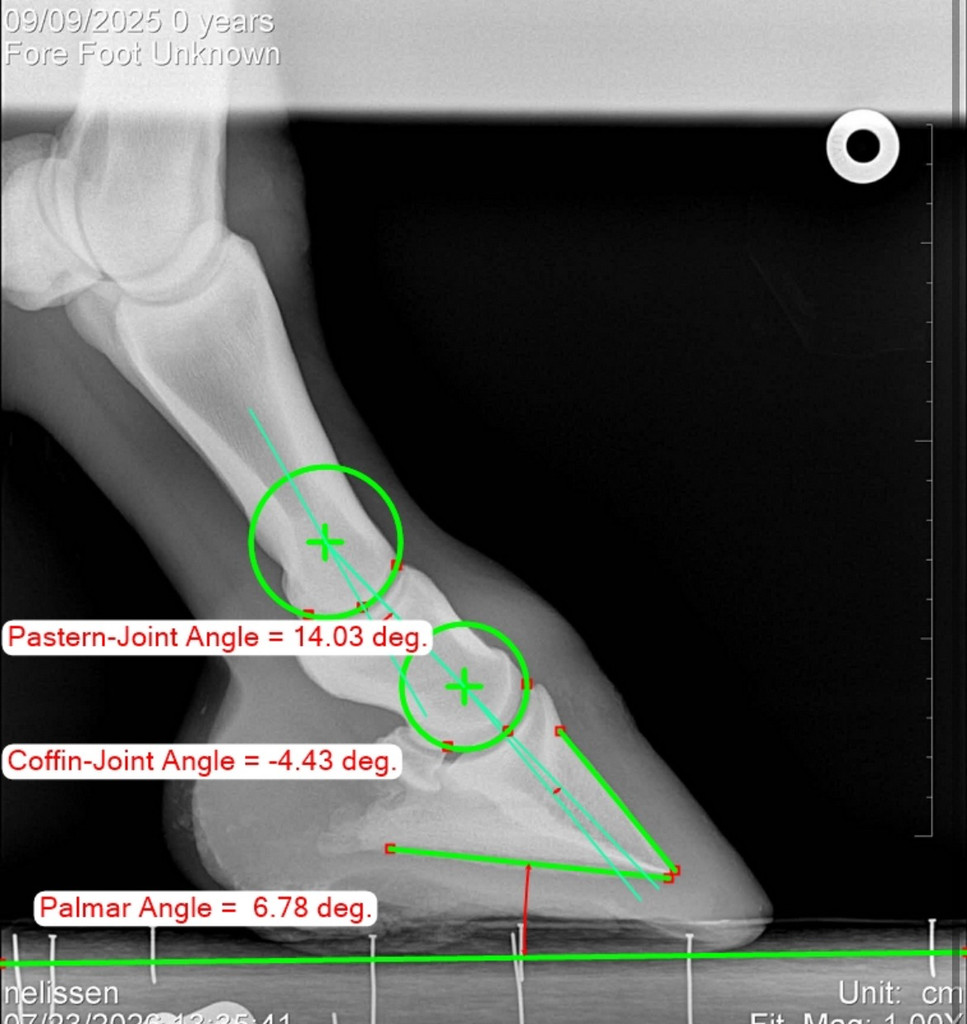
A radiograph might be able to suggest to us that a horse might benefit from something like boots and pads for protection, if we see thin soles or issues with internal structures, but that still doesn’t tell us the entire story.
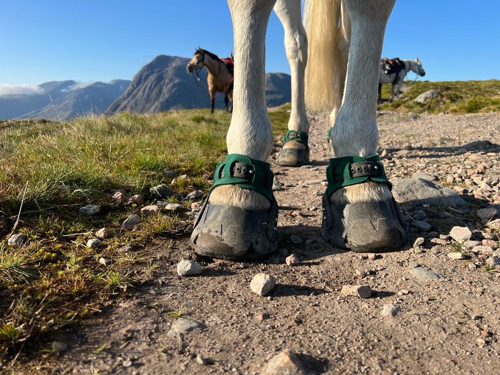
A Radiograph Can’t Tell Us What Hurts
A radiograph can show an abnormality, but it cannot necessarily tell us whether that abnormality is currently painful. Those aren’t the same thing.
Think about how often this happens in human medicine. Someone has imaging performed for one reason and the doctor happens to discover arthritis, a bone spur, evidence of an old injury, or another structural change that the person never even knew existed. They weren’t painful there. They weren’t having a problem. It was simply an incidental finding.
The same thing can happen with horses. We can find remodeling or abnormalities on a radiograph that look significant and naturally assume that must be why the horse is lame. Maybe it is, but the radiograph alone doesn’t prove that. Some radiographic changes may have been present for years without causing the horse any obvious discomfort.
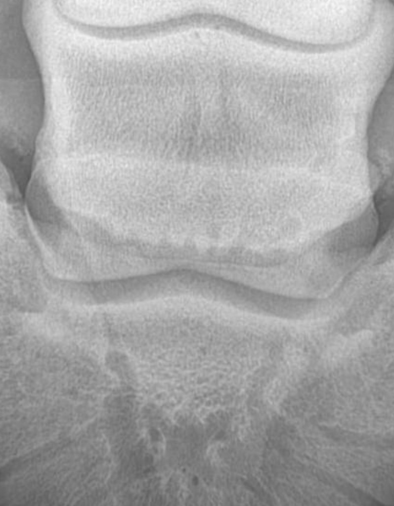
This is why the clinical picture matters so much. We have to look at where and when the horse is uncomfortable, how they move, whether the lameness changes on different surfaces, what happens on a circle versus a straight line, what the veterinarian finds during a lameness examination, and, when appropriate, what happens with diagnostic nerve or joint blocks. The imaging finding needs to correlate with the horse in front of us. Otherwise, we risk treating an image instead of treating the horse.
Radiographs Can’t Show Us Everything Inside the Hoof
Another important limitation to radiographs is that the hoof contains much more than bone. There are tendons, ligaments, cartilage, blood vessels, nerves, the digital cushion, laminae tissues, and numerous other soft tissue structures packed into a relatively small area.
Standard radiographs are excellent for evaluating mineralized structures, but they don’t allow us to directly evaluate most of these soft tissues in the same way. For example, the deep digital flexor tendon travels down the back of the limb, through the navicular region, and attaches to the coffin bone. A horse can have significant pathology involving that tendon without the injury itself being clearly visible on a standard radiograph. The same is true for a number of other soft tissue injuries within the foot.
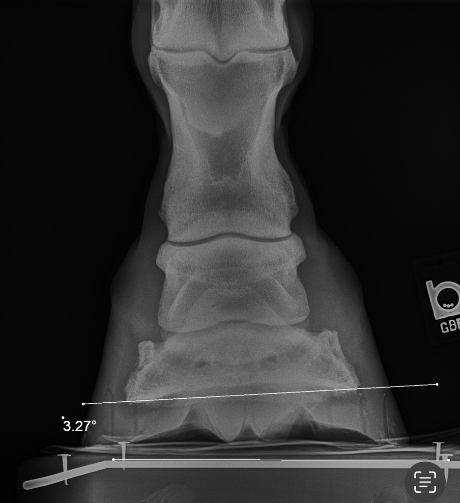
Sometimes radiographs provide indirect clues that make a veterinarian suspicious of what’s happening around a particular structure, but that is very different from actually visualizing the soft tissue injury itself. Depending on the suspected problem, additional diagnostics such as ultrasound, MRI, or bone scan may provide information that radiographs cannot.
This also means that a horse having “clean” radiographs doesn’t necessarily mean that nothing is wrong. It simply means that the radiographs did not reveal a bony or mineralized abnormality that adequately explains the horse’s symptoms.
Bone Changes Don’t Automatically Equal Pain
Bone responds to stress throughout a horse’s life. It remodels, adapts, and can retain evidence of previous injuries or loading patterns. Because of this, especially as horses age, we may see things on radiographs that don’t look textbook perfect.
When we find something on radiographs, we have to ask, “Is this finding clinically relevant to what this horse is experiencing right now?”
Sometimes the answer is relatively straightforward. If a horse suddenly becomes severely lame and radiographs reveal a fracture in the area that has been localized as painful, there may be a very strong connection between the imaging and the clinical signs. Other findings aren’t nearly as clear.
This is where we have to be careful about allowing an image to become a diagnosis all by itself. An abnormality deserves consideration, but it also needs context. The horse’s history, examination, movement, response to diagnostic analgesia, and potentially other imaging all help determine whether what we’re seeing on the radiograph actually matters to that horse.
What About All Those Angles?
Radiographs also allow us to measure angles and relationships that we can’t accurately determine by simply looking at the outside of the hoof. This can be incredibly helpful, but those measurements can also become overinterpreted.
We might see a negative palmar angle, a long toe, decreased sole depth, an unusual phalangeal alignment, or another measurement that differs from what we’d consider ideal. That gives us information, but a measurement isn’t automatically a diagnosis, and it doesn’t necessarily tell us why that relationship exists.
We still need to consider the horse’s individual conformation, hoof capsule distortion, heel position, toe length, sole depth, bone remodeling, and the rest of the limb. We also need to consider how the radiograph itself was taken. The horse’s positioning, how evenly they were standing, beam placement, the block beneath the foot, and other technical factors can influence what we see and measure.
Numbers are useful. They can give us objective information and help us track changes over time. But they still have to be interpreted within the context of the individual horse.
Radiographs Don’t Tell Us Exactly How to Trim the Horse
Some people want to use radiographs to determine how to trim a horse. There is a little problem with that, though: not all horses read the “correct hoof” textbook.
Radiographs can absolutely provide a hoofcare professional with valuable information. They can help us better understand sole depth, internal hoof orientation, hoof capsule distortion, and the relationship between the coffin bone and the external hoof. That information may influence our trimming decisions, but it doesn’t automatically dictate them.
For example, let’s say a radiograph shows what appears to be a long toe. That doesn’t automatically mean we should aggressively remove everything that appears to extend beyond some predetermined measurement. We still need to look at what tissue is actually available to safely remove, how much sole depth the horse has, the quality of the hoof wall and sole, the horse’s conformation, how the horse is loading the foot, how they’re landing, and most importantly, how comfortable they are.
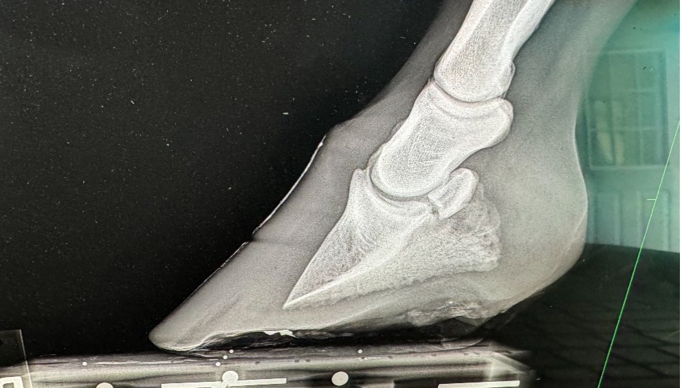
The same is true when we see an angle on a radiograph that we don’t like. Our goal shouldn’t necessarily be to make that angle look “normal” as quickly as possible. If the hoof capsule developed its current shape over months or years, dramatically changing the foot in one trim may not be appropriate or comfortable for that horse.
Sometimes the best response to what we see on a radiograph is actually patience. We may make a conservative change, allow new hoof to grow, see how the horse responds, and reassess over subsequent trim cycles. A horse’s response to those changes gives us additional information that the original radiograph could never provide.
Radiographs Are a Snapshot
One radiograph gives us a picture of what was happening at one moment in time. Serial radiographs can sometimes give us something even more useful: a story.
When images are taken using consistent positioning and technique, we can compare them over time. This can be particularly valuable when monitoring laminitis, significant hoof capsule distortion, sole depth, bone remodeling, or complicated rehabilitation cases.
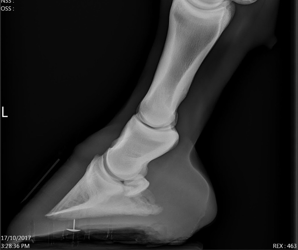
Instead of simply asking whether today’s radiograph looks “good” or “bad,” we can look at what is changing. Is sole depth improving? Is the relationship between the hoof capsule and coffin bone changing? Is bone remodeling progressing? Are structural changes stabilizing? And, just as importantly, do those radiographic changes correlate with changes in the horse’s comfort and movement?
This is also why good radiographic technique matters. If we’re going to make very small measurements or compare images taken months apart, positioning needs to be as consistent as possible. A few degrees of apparent change may look exciting or alarming, but if the horse was standing differently between the two images, we need to be cautious about what conclusions we draw from that difference.
The Horse Still Matters More Than the Picture
I love having radiographs available on complicated hoof cases. They can answer questions that we simply cannot answer by looking at the outside of a foot. They can change the way we approach a case, alert us to something requiring veterinary attention, and provide incredibly useful information when veterinarians and hoofcare providers are working together.
But we never want to forget the horse attached to those images. A horse isn’t a coffin bone floating inside a hoof capsule. There are tendons, ligaments, joints, nerves, connective tissues, vascular structures, and an entire limb above that hoof. There is a nervous system interpreting pain. There may be metabolic factors affecting tissue health. There is a history behind that horse, an environment they’re living in, and a unique way that they move and load their feet.
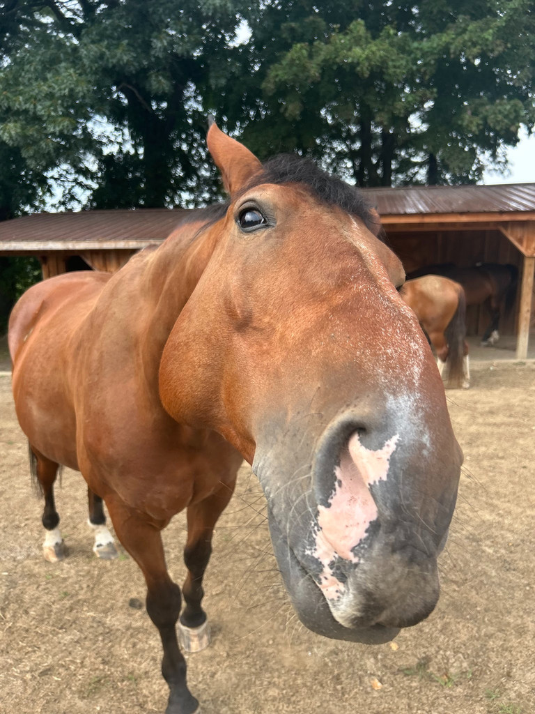
Radiographs give us incredibly useful information about structure, but they cannot independently tell us about pain, function, or cause. And they certainly can’t hand us a universal trimming prescription.
The best use of radiographs is to combine what we see on the image with the veterinary examination, the horse’s history, movement, hoof assessment, other diagnostics when necessary, and the horse’s response to what we do.
We don’t want to just make a radiograph look perfect – we want to help the horse become – and remain – as comfortable and functional as possible.